What They Had is a 2018 movie focusing on the family whose matriarch suffers from Alzheimer’s disease. Although seemingly similar, this movie is very different from the more popular Still Alice, and, in my opinion, shows the difficulties of navigating everyday life better. The movie, directed by Elizabeth Chomko, who drew inspiration directly from her personal life, stars brilliant Hilary Swank as a worried daughter Betty who comes back home in order to care for her mother Ruth, played by Blythe Danner. Michael Shannon, playing Swank’s brother Nicky, is masterful as ever, showing his impeccable acting skills, as he clashes with their father (Robert Forster) over the possibility that their mother should continue her life in an assisted living facility.
What I like about this movie, is that it isn’t solely focused on Alzheimer’s disease, but rather on family dynamics around it; it explores how members of the immediate family deal not only with their mother, wife, and grandmother as the illness progresses but also with each other. Without revealing more of the plot, as I truly believe everyone should watch this movie, I have to praise it for tastefully and realistically conveying many symptoms of AD, such as when Ruth wanders away in the middle of the night, experiences speech difficulties or flirts with her adult son, not recognizing him.
The last scene I described isn’t brought on as a dramatic one, but rather a humorous one, albeit dark. As a matter of fact, the whole movie deals with the severity of the situation in a similar manner, but it will still bring tears to your eyes. This movie is honest, truthful, and above all, touching; I honestly can’t believe it’s not more known and as I’m writing this review, I’m quite anxious that my review isn’t presenting it in the best possible light, which it absolutely deserves. I hope my clumsy writing won’t discourage you from watching What They Had, a movie that beautifully reminded me why I chose neurodegeneration as my field of study.
If you watched What They Had, let me know if you liked the movie as much as I did. I am also sharing the trailer here as well.
Phylum Platyhelminthes, also known as flatworms, consists of four distinct classes: Turbellaria, Monogenea, Trematoda (flukes), and Cestoda (tapeworms).
Today, I want to write more about Turbellarian nervous system, which is more advanced than the one found in Ctenophora or Cnidaria. Turbellaria are small animals (up to 20 mm in size, although there is one species that can be more than half a meter long, imagine that touching your foot) found in water and wet habitats. Turbellaria have a brain, both sensory and motor neurons, and a series of sensory receptors. Although bilateral animals, not all Turbellaria have a bilateral nervous system, with some of them still having a radial system characteristic for cnidarians.
The turbellarian nervous system, made of uni-, bi-, and multi-polar neurons, can be epidermal, sub-epidermal, and sub-muscular. However, only less advanced species have the epidermal nervous system, while all the others have both subepidermal and submuscular.
When discussing the radial nervous system, it is important to mention cerebral ganglion and three pairs of nerve cords (dorsal, lateral, and ventral). These cords are connected by annular commissures. In the bilateral system, on the other hand, we have a primitive brain made of several ganglia and only two ventro-lateral cords, mutually connected by transverse commissures. There are also sensory nerves, which extend forward from the brain.
Turbellaria have a whole myriad of sensory receptors: mechano-, chemo-, photo-, and balance receptors. Mechanoreceptors can be divided into two groups, thigmoreceptors and rheoreceptors. Both of these can be found on the whole area of Turbellaria body, and both contain cilia in order to sense outside stimulus. The difference between the two is that thigmoreceptors are specialized for touch, while rheoreceptors process water flow stimuli. Chemoreceptors are located in special grooves on the head, and serve to locate food or a mate. Photoreceptors are located in ocelli (ocelli are analogue of eyes) and although they usually have only a pair on the head, some species have couple of pairs or even many ocelli on the edge of their bodies. Statocysts serve as balance organs, although only some species have them. Statocysts are chambers filled with a fluid and also contain one statolith. It is actually unknown how statocysts receive stimuli.
Schmidtea mediterranea, an adorable flatworm of Tricladida class is one of the modal organisms in genetic and molecular research, because it has diploid genome and asexual and sexual strain. These characteristics make S. mediterranea a very popular choice among the scientists, especially since the discovery of its apparent immortality. Due to an abundancy of stem cells, almost any amputated part of this flatworm can regenerate into a full organism in a span of just several days. Yes, this little organism is literally Deadpooling its way through life!
Of course, the explanation behind this mechanism is all but simple; it seems that this regeneration ability depends much on the activity of an enzyme called telomerase, and not even this works the same in asexual and sexual strains of S. mediterranea. There are also many genes involved, but since some of the genes have orthologs in human, scientists are now trying to discover if they could somehow stop aging in our species.
So, what do you think about these small creatures? Do you like them, or do they frighten you a little bit?
Unfortunately, I couldn’t find many resources online regarding their general nervous system, so most of the information is sourced from one book, which is available only in Croatian. More research is needed regarding these creatures, and some are underway, especially regarding their astonishing regenerative capabilities.
Literature & more information: Habdija et al: Protista-Protozoa, Metazoa-Invertebrata, Alfa, 2011, Zagreb Moraczewski, Czubaj & Bgkowska Organization and Ultrastructure of the Nervous System in Catenulida (Turbellaria) Zoomorphologie 87, 87-95 (1977) Tan et al: Telomere maintenance and telomerase activity are differentially regulated in asexual and sexual worms PNAS vol. 109, no. 11, 4209–4214 (2012) Handberg-Thorsager, Fernandez & Salo Stem cells and regeneration in planarians Frontiers in Bioscience 13, 6374-6394 (2008)
Ctenophora, commonly known as comb jellies, are a rather perplexing phylum of beautiful pelagic creatures. Their evolutionary position has been debated for many years as is the origin of their nervous system (some scientists believe they are older than sponges and that sponges lost their nervous system, while others advocate the theory about the nervous system forming independently twice, once in cnidarians and once in ctenophores). Ctenophora have two nerve nets: subepidermal and less organized subgastrodermal, which recent research identifies as a mesogleal nerve net. Nerve cells from this layer communicate with muscles by synapses and affect the locomotion of the body. The subepidermal net is denser around the mouth, the pharynx, and under the comb rows (comb rows are strips that run the length of the ctenophore body and contain cilia called “ctenes”). Ctenophore neurons can be iso- and multipolar.
They have sensory cells on the whole surface of the body and those correspond to vibrations and thermal and chemical stimuli: more receptors are located around the mouth and pharynx. Ctenophora also have an apical and aboral sensory organ. Such sensory organ consists of a statocyst, a sensor that contains a statolith that balances on four groups of long cilia connected to the comb rows. These organs help the orientation of the ctenophore body. What’s extremely interesting is that ctenophores use different chemical signalling system than the ones described in the previous posts, mainly because these animals simply lack the neurotransmitters (and genes), such as serotonin, dopamine, noradrenaline, and acetylcholine; glutamate is the only neurotransmitter currently known to be present.
I gathered all this information from different resources, and some are sometimes contradictory or are generalizing conclusions about the whole phylum from the data of only one ctenophora species. This is the best overview I could manage, to show both the similarities and the differences of the ctenophora nervous system, when compared to the Cnidarian system. These lovely animals are not very well researched and I’m sure many wonderful breakthroughs about their anatomy, physiology, and their place in the evolutionary tree are to come.
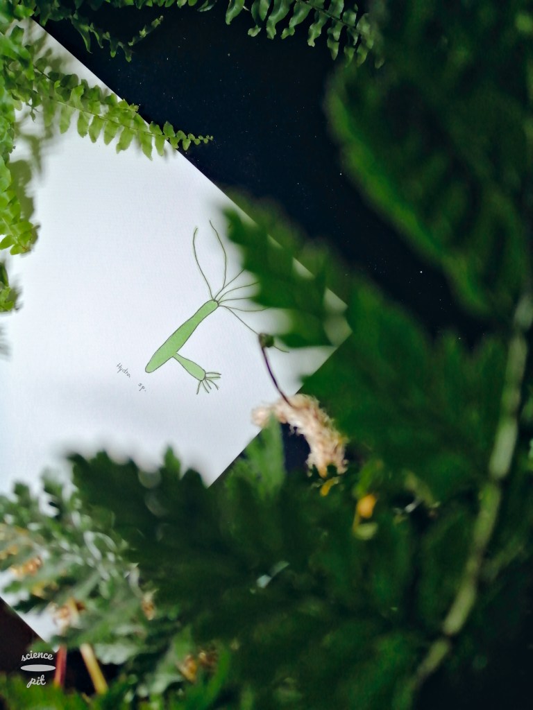
Hydrozoa are the last cnidarian class I’m going to write about. They can exist in two distinct shapes, as hydromedusa and hydropolyp (same as Scyphozoa and Cubozoa). Despite perhaps expecting hydrozoans to be the most advanced in both nervous and sensory systems, they don’t actually have any rophalium. Furthermore, some hydromedusae don’t even have nerve nets. However, they have two nerve rings (outer and inner) on the margins of their bells which are regarded as ganglia by some scientists.
These rings consist of neural pathways which process different sensory inputs (such as light and gravity). Aglantha digitale, a hydrozoan species, has been reported to have as much as 14 distinct neural pathways. A. digitale is also distinct from the other species in the class by having two swimming “modes” – slow (which is a characteristic for all hydrozoan) and escape mode. Transmission through giant ring neurons is responsible for both modes, but the escape mode requires a stronger contraction. The slow swim mode is activated by the input from the pacemaker, which triggers slow calcium spikes. Direct mechanical nerve ring stimulation by tentacles triggers fast sodium spikes. In short, giant ring neurons are capable of generating two different kinds of action potentials.
bsh
Gap junctions are also present in (and only in) Hydrozoa, and they transfer electrical signals through the musculature. Furthermore, I would like to emphasize that despite some hydromedusae not having a nerve net, some in fact do, and so do hydropolyps. In polyps, however, some groupings of the neurons could be found around their mouth.
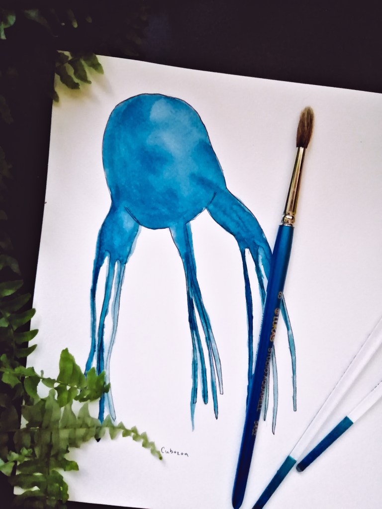
Cubozoa, or box jellyfish, are another cnidarian class. Their name stems from their distinct cube-like shape. Cubozoa are also distinct from other cnidarian because their venom can be fatal to humans. As with all cnidarians, box jellyfish have two nerve nets and, like Scyphozoa, rophalia. However, box jellyfish also have a distinct nerve ring, as well as more developed eyes that consist of a lens, cornea, pupil, and a layer of retinal cells. Altogether, Cubozoa have 24 eyes, which makes them the most advanced cnidarian class in the sensory aspect.
Rophalia are mutually connected via the mentioned nerve ring. This ring is believed to be an integration center for the swimming, visual, and tentacle systems; it is comprised of oversized neurons, as well as some smaller neurites. The communication between the nerve net and jellyfish muscles is regulated by chemical synapses.
Most of the information relating to Cubozoa, I already mentioned in the previous post about Scyphozoa, so I only wanted to relay the main differences between the two. These two classes are so similar that, until recently, they were actually considered one class.
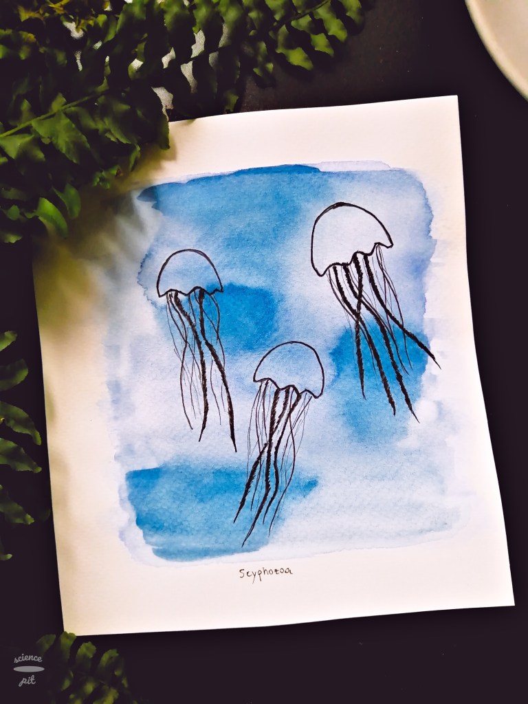
Scyphozoa (true jellyfish) are much more interesting (in a neurobiological way) than previously described corals. One major difference is that Scyphozoa are pelagic animals, which means they are not fixed to the ground. They also have two diffuse nerve nets (subepidermal and subgastrodermal) that consist of bipolar and multipolar neurons – the impulse conduction has been measured at 0,15 m/s. Both nets coordinate the movements of an animal towards the food. Some scientists, however, differentiate one diffuse and one motor nerve net. The motor net is in charge of the activation of muscle contractions after receiving signals from the so-called pacemaker organs (which are in charge of the swimming rhythm). The diffuse net, in this case, is in charge of marginal tentacle contraction and it is also believed it communicates sensory information to jellyfish musculature. Neurons of the motor nerve net are connected by chemical synapses, while neurons of diffuse nerve net are connected by peptidergic synapses that were noted in Anthozoa as well.
Sycphozoa also have much more developed sensory organs than any of the animals previously mentioned. These sensory structures are called rhopalia and they are located on the edges of the jellyfish bell – there are usually four of them (or a number that’s a multiple of four). Rhopalia contain multiple sensory receptors – statocyst (balance receptor), ocelli (light sensitivity), a mechanoreceptor, a chemoreceptor, and aforementioned pacemaker neurons.
I would also like to note here that some authors (I’m referring here to the article “Do jellyfish have central nervous systems?” by R. A. Satterlie) believe this kind of nerve net explanation is rather simplified and that there exist some evidence suggesting that jellyfish have a centralized nervous system, mainly that rophalia are in fact rudimentary ganglia and could be regarded as integrative centers. However, any communications between rhopalia themselves exist only through the nerve nets.
I don’t know about you, but I just love doing online courses, especially when they deal in subjects I don’t get to explore in my college courses. Over the years, I tried many different platforms, such as YouTube, Google Digital Garage, Khan Academy, Udemy, and, my favourite, Coursera. As a matter of fact, I discovered Coursera back when they started in 2012; most of the courses I took were on topics of Neuroscience and Molecular Biology. At first, courses and certificates were completely free, but with time, they started offering paid vs. free, as well as many specializations and even some college degree courses. However, many of the courses are still available to watch and do quizzes, just without the certification.
Disclaimer: this post is not sponsored by Coursera.
This course, offered by Hebrew University of Jerusalem, is actually the very first course I took, and at the time was one of the rare Neuroscience courses. I have only good memories about this one – it is a good introductory course into the field and the professor explained the curriculum very well. Also, the course mentions real projects that deal with neural networks and brain reconstructions, such as Blue Brain Project. I didn’t mention this previously, but every course also comes with subtitles (in English at least) and transcripts, so you can follow along easier.
The Addicted Brain (by Emory University) is another course appropriate for beginners in this topic – I was initially interested not only because of the topic of addiction, but also because various mechanisms of how drugs interact with the brain were presented. The course also covers the topic of drugs in society, although this part mainly concerns United States of America. Also, I don’t know if this is something that’s important to you, but I followed professor’s narration easily – his voice is calming and he speaks very understandably.
Medical Neuroscience (by Duke University) is not only the most advanced course of the ones mentioned here, but the most advanced course I ever took. Actually, I started it once or twice before, but dropped out because it required a lot of time and dedication that, at times, I just didn’t have due to my University obligations. This course is really extensive and requires some before-knowledge, but is also very satisfactory when you finish it. The only problem I had with this one is that sometimes I felt that questions in quizzes were asking for details that to me seemed almost overlooked in the videos. However, I felt like this course was quite important for my studies, since I have a strong interest in Neuroscience, but lacked the medicinal perspective.
All quizzes are multiple choice answers, with usually one correct answer (sometimes more correct answers). I vaguely remember some questions where you had to connect some phrases (like 1-d, 2-c, etc) as well, but haven’t came across those recently. Also, the quizzes I did were never timed and you can take one quiz 3 times every eight hours (they keep your highest score).
Coursera also offers financial aid – you can fill out an application where you explain why is the course you’re applying for important to you and why you can’t afford it. So far, I’ve heard of many positive experiences where they gave grants.
There are also two Neuroscience related courses I am planning to take – Human Neuroanatomy (to revise a bit) and Computational Neuroscience, which deals with using Python in Neuroscience research. I would very happily review those for you, in a greater detail, if this is something you’d like to read about!
***
What is your opinion on online courses – do you think they’re useful or a waste of time? Did you perhaps take some of the ones I mentioned? If yes, I would love to hear from your!
Anthozoa (corals & anemones) are interesting animals that live exclusively in seas and oceans. Since they are regarded as sessile organisms, their nervous system is not very advanced. In a previous post, I mentioned that all cnidarians have two diffuse neural nets, and that is true for Anthozoans as well. Multipolar nerve net neurons are connected with synapses, and they also possess sensory cells that are particularly numerous around the mouth and on the tentacles.
One paper investigated traveling of electrical waves in a coral nerve network in coral colonies in genus Palythoa. It was experimentally observed that this electrical wave spreads at the constant speed from the site of simulation. Furthermore, peptidergic neurons (the ones using neuropeptides to communicate) were also noted in Anthozoa.
Did I draw this when I was 5 or 25? – A mystery.
Nematostella vectensis, also known as starlet sea anemone, is a species of Anthozoa that is known as a model organism – its genome and development have been carefully studied, including its nervous system. In this species, oral and pharyngeal nerve rings have been reported, as well as longitudinal tracts of neurites (neurites are usually axons or dendrites). These findings would suggest that some groupings of neural cells exist in at least some Anthozoan species after all. Sensory neurons, interneurons, motorneurons, and neurosecretory‐like gland cells were also reported to exist in N. vectensis.
Note: in case I didn’t mention this before, cnidocytes are often considered neural cells because they display mechanosensory properties and calcium dependent neural‐like properties as well.
A nerve net is a type of nervous system that consists of many neurons but there is no brain or cephalization. Nerve nets are found in animals with radial symmetry (Cnidaria) and biradial symmetry (Ctenophora). Despite being called a net, there sometimes exist some groupings of neural cells in some Cnidaria classes, which I will write more about during the next couple of weeks. Cnidaria are specific due to their specialized organelles, cnidocytes, which they utilize to hunt for food or use for securing itself to a surface. Some cnidocytes contain toxins that can paralyze their prey (the burning sensation you may have felt when touching a sea anemone 😉). As a rule, Cnidaria have two diffuse nerve nets, one in the epidermal layer and a second one in the gastrodermal layer. In between these two layers is the mesoglea, a layer that functions as sort of a skeleton. The epidermal net consists of bipolar and multipolar nerve cells, while the gastrodermal net is made up of only multipolar cells.
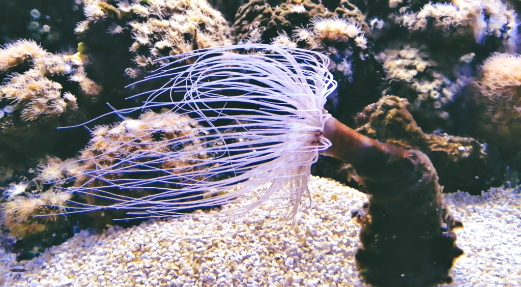
Cerianthus membranaecus (known as cylinder anemone or coloured tube anemone)
Cnidarian nerve systems are fascinating but also quite unexplored. What is known is that nerve cells consist of two types of neurons, sensory neurons that respond to stimuli and motor neurons which ultimately trigger a response. Chemical synapses exist and provide the communication between the neurons. Hormones have also been reported in some cnidarians (steroids, neuropeptides) but it is still not known how exactly these signalling molecules work.
***
In the next couple of weeks, I will write a post about every cnidarian class and also ctenophores, focusing on their nervous and sensory systems. If you have any questions or would like me to focus on something, please let me know!
Sponges (phylum Porifera) are sessile multicellular organisms that live predominantly in seas and oceans. They don’t have tissues or organs, and therefore, they don’t actually have a nervous system. However, they do have bipolar and multipolar cells that resemble nerve cells, which are found in the middle, “jelly-like”, layer. Sequencing of some sponge species showed the presence of many genes associated with neural cells, such as genes that code enzymes for neurotransmitter synthesis and synaptic transmission. It is important to note that these genes have other functions in the organism. It has also been observed that some sponge larvae can respond to outer stimuli and show various “taxis” behaviour – phototaxis (response to light), geotaxis (response to gravity), rheotaxis (response to water current). Phototaxis has been closely studied in species Amphimedon queenslandica (class Demospongiae), a sponge native to Coral Sea.
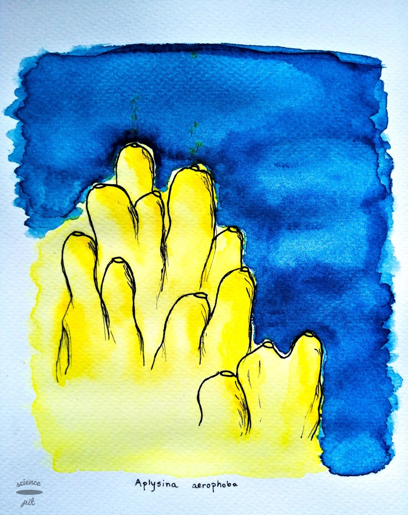
Aplysina aerophoba, also of class Demospongiae, which can be found in Adriatic Sea.
Potassium channels have been observed in that same species, as well as glutamate, GABA, and NO systems, which have been investigated in Ephydatia muelleri, another species of class Demospongiae. Electrical signalling has been noted in glass sponges (class Hexactinellida). These sponges have bodies comprised of a syncitial tissue and their skeleton is made of silicon dioxide. The scientists were able to measure the action potential (5s long, with 29s refractory period) and deduce this signal relies on potassium and calcium ions. Some scientists even suggest that sponges used to have a nervous system, but lost it during evolution – they introduced several hypothetical scenarios for this event, proposing that sponges lost their nervous system in order to focus on filtering.